An article on the cost of endoscopy test in Nigeria, diagnostic and therapeutic endoscopy, Upper GI endoscopy, lower GI endoscopy, preparing for endoscopy, Endoscopic Retrograde Pancreatography (ERCP).
This article will also discuss the pricelist of endoscopy in Nigeria, endoscopy test cost in Nigeria and endoscopy price in Nigeria depending on how you search it on Google.
FACTS ABOUT ENDOSCOPY
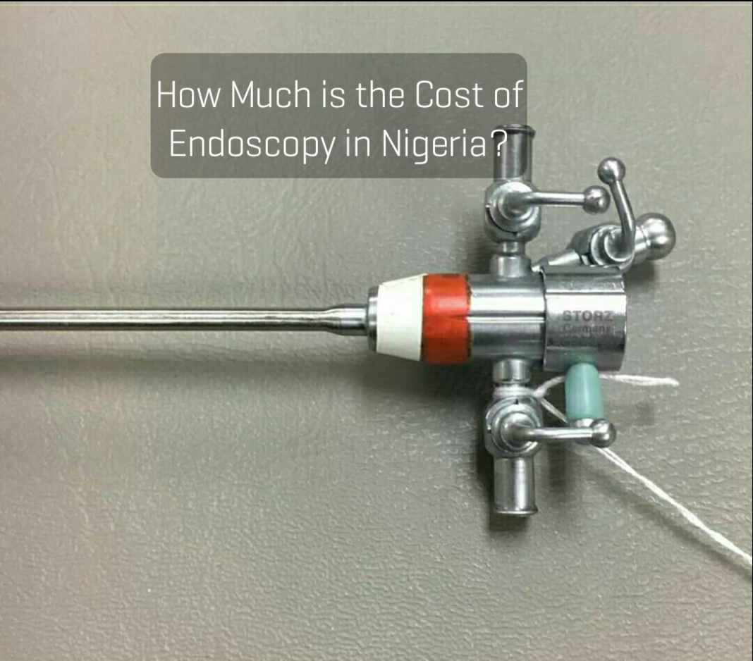
➡️Endoscopy is the direct visualisation of the interior of organs and structures, using either rigid or flexible instruments.
➡️The development of fibreoptic endoscopes has made possible the examination of distal structures not accessible to the rigid scope.
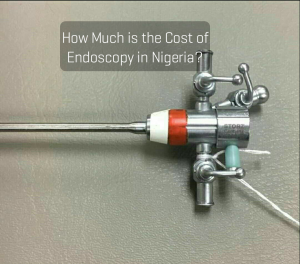
➡️Flexible endoscopy has the range and applicability of pre-operative diagnostic techniques, intraoperative endoscopy and postoperative follow-up.
➡️Endoscopy has evolved to be a versatile tool in surgical practice, both for diagnosis and treatment and follow-up care. Treatment can sometimes be effected at the same time that diagnosis is made.
➡️IT can be safely carried out on outpatient basis. You will need only be given a local anaesthetic or sedated with small amounts of benzodiazepines/narcotics (the commonest drug used is Midazolam) to calm you down and reduce your anxiety.
➡️The sedated patients are, however, be monitored and cared for to minimise and manage the side-effects of the drugs used.
➡️The services of an anaesthetist may be useful in some patients, especially in children and those with pre-existing cardiorespiratory or hepatic disease.
➡️The cost of endoscopy test in Nigeria depends on the diagnostic centre, the doctor and the type of endoscopy to be carried out.
TYPES OF ENDOSCOPY
DIAGNOSTIC ENDOSCOPY
💥As the name implies,it is done to make diagnosis. This provides for direct visualisation of lesions, especially those in which indirect methods of imaging are not so accurate.
💥It is particularly useful in cases of gastro-intestinal bleeding and visualising suspicious lesions found on contrast x-ray examination.
💥It provides an opportunity for biopsy (sample of your tissue taken for test) in many situations without resort to an open procedure.
💥Apart for the benefits stated above, diagnostic endoscopy permits for the use of endosonography for the pre-operative staging of such lesions as rectal and oesophageal cancer.
💥Endorectal ultrasonography is also valuable in the evaluation of prostatic disease, especially in localising areas in the prostate for biopsy,bronchial washings can also be done at bronchoscopy and the fluid sent for cytological examination.
THERAPEUTIC ENDOSCOPY
Therapeutic endoscopy is done to achieve cure and relieve symptoms. It permits the use of the following treatment methods:
1. Mechanical dilatation of stenotic lesions (e.g.oesophagcal strictures) using bougies or balloons.
2. Placement of stents.
3. Injection sclerotherapy
4. Rubber band ligation, eg. for oesophageal varices
5. Electrocautery
6. Laser ablation
7. Placement of feeding tubes
8. Extraction of foreign bodies
9. Instillation of treatment agents, e.g.for gallstone dissolution.
10.Papillotomy/sphincterotomy for extraction of common bile duct stones.
11. Resection of growths and tumours-e.g. enlarged prostate and bladder tumours.
Apart from the upper and lower gastrointestinal tract, these procedures can be done in the trachea and bronchi (bronchoscopy),the joints (arthroscopy), and urinary tract – urethrocystoscopy or ureteroscopy.
Upper Gastrointestinal (GI) Endoscopy (Oesophago-Gastro-Duodenoscopy, OGD)
💥Rigid endoscopes are available for the visualisation of the oesophagus and for extraction of foreign bodies – under general anaesthesia. Flexible scopes, however, are less traumatic and can be manoeuvred to reach the duodenum.
💥In the oesophagus, upper GI endoscopy can be used to investigate the aetiology of dysphagia (difficulty in swallowing) caused by inflammatory conditions (moniliasis is sometimes seen in patients who are immune compromised e.g. HIV), obstructive lesions that block the passageway (tumours, foreign bodies, food impaction, etc), strictures (benign or malignant), or functional motility disorders (achalasia, etc).
💥Brushings or biopsies can be taken (from tumours for definitive diagnosis and has a far superior pick -up rate than that of barium X-ray examination.
💥Strictures or obstructive tumours may be dilated to provide some relief of symptoms.
💥Pneumatic balloon dilatation may also be useful in achalasia. Placement of
stents may provide short-term palliation of symptoms from inoperable malignant strictures.
💥Laser ablation of tumours is another treatment option. The injection of botulinum toxin into the hypertensive lower oesophageal sphincter (LOS) leads to decreased LOS pressures, and may provide short term relief of symptoms. This is a useful option for patients who have a poor risk for operative therapy.
💥In upper GI bleeding, early endoscopic evaluation helps to differentiate between bleeding oesophageal varices and bleeding peptic ulcers, Mallory-Weiss tears, benign and malignant tumours and more uncommon vascular lesions like the Dieulafoy lesion and angiodysplasia.
READ ALSO: A to Z of Liver Function Tests.
💥Visualisation of a bleeding peptic ulcer has some predictive value. Bleeding is likely to continue or recur with the following stigmata: visible vessel in the ulcer base, arterial bleeding or oozing and a clot overlying the ulcer.
💥Early operative intervention is advisable in these situations, especially if there is cardiovascular instability.
💥Varices can be treated endoscopically by banding or injection sclerotherapy. Bleeding peptic uleers can also be treated endoscopically.
💥Gastric and duodenal lesions can also be visualised and biopsied. Biopsy specimens are subjected to histopathology to confirm diagnosis of benign or malignant tumours. Multiple biopsies are always taken from the inner edge of ulcers in the stomach because or the risk of malignancy (cancer) in gastric ulcers.
💥Mucosal biopsies may show gastritis (inflammation of the stomach) with or without Helicobacter pylori infcction, metaplasia or dysplastic changes.
💥Other therapeutic procedures that can be carried out include drainage of pancreatic pseudocysts and inserting percutaneous endoscopic gastrostomy (PEG) tubes.
Preparation for Oesophago-Gastro-Duodenoscopy (OGD)
Patients are instructed to fast for six hours to ensure that the stomach is empty.
If you have gastric outlet obstruction, a flexible pipe will be placed in your stomach through your nose by your nurse or doctor to empty it and gastric washout is done to clear food debris in the stomach before OGD is undertaken.
Patients need-to be informed about what the procedure entails to ensure adequate co-operation.
How upper GI Endoscopy is done
💥The patient’s throat is anaesthetised with a topical spray (e.g. xylocaine) or is sedated with small amounts of a benzodiazepine.
💥Patients who are sedated should leave hospital accompanied and should not drive, use machinery or drink alcohol for the rest of the day.
💥During the procedure, the patient’s pulse and oxygen saturation should be monitored by a pulse oximeter where facilities arc available .
💥The procedure is carried out in the left lateral position and care taken to avoid aspiration of secretions and gastric contents.
💥Patients whose throats are sprayed for the procedure are allowed to drink only when the anaesthetic wears off, usually
within two hours.
Endoscopic Retrograde Cholongiopancreatography (ERCP)
💥ERCP is a useful technique in examining the duodenum and ampulla of Vater, and outlining the pancreatic and bilary ductal system. Endoscopic visualisation and appropriate biopsies can achieve diagnosis of periampullary carcinoma.
💥Abrupt ductal obstruction and irregularity on ERCP wiII corroborate the clinical and othcr evidence of pancreatic carcinoma.
💥Calculi (stones)in the biliary tract and other strictures will be evident from contrast examination.
💥ERCP is therefore an invaluable investigation of obstructive jaundice.
ERCP can be used therapeutically in the following settings:
1. Extraction of common bile duct (CBD) stones, before or after cholecystectomy.
2 Dilatation of strictures using hydrostatic balloons.
3. Placement of slents to maintain patency in obstructive lesions of the CBD.
4. Planning operative strategy if surgery is required following an abnormal cholangiogram.
Lower Gastrointestinal Endoscopy
Endoscopy of the lower gastrointestinal tract is useful for diagnosis, treatment and surveillance of anal and colorectal
lesions. It is also called colloquially as colonoscopy.
The instruments used are the proctoscope, the rigid and flexible sigmoidoscope and the colonoscope.
Lower GI endoscopy can therefore be classified under:
➡️Proctoscopy
➡️Sigmoidoscopy,
➡️Rigid sigmoidoscopy
➡️Flexible sigmoidoscopy
➡️Colonoscopy
➡️Proctoscopy is a useful adjunct to digital rectal examination and is the procedure of choice in examining and treating anal lesions, e.g. haemorrhoids. It is performed at the outpatient clinic and needs no prior preparation.
It is contraindicated when there is severe anal pain, e.g. in anal fissures.
The rigid sigmoidoscope can also be used at the out patient clinic although views maybe somewhat compromised by faeces in the unprepared bowel. It is difficult to manoeuvre the instrument proximal to the sacral promontory and so the range of vision is limited.
Flexible sigmoidoscopy uses the fibreoptic flexible scope and may therefore be manoevred much further.
Like colonoscopy, the bowel must be prepared i.e. emptied of faecal contamination.
Colonoscopy permits visualisation of the entire rectum and colon, and its use can be extended to visualise the distal 20cm of the ileum.
The patients must be given analgesia or sedation and the procedure requires the skill of an experienced endoscopist.
The proctoscope and rigid sigmoidoscope can be advantageously utilised in certain situations. They:
1. complement digital rectal examination (DRE).
2. can be used in the unprepared bowel, or with little bowel preparation.
3. permit visualisation of the anus and rectum in a manner that is difficult to achieve using the flexible sigmoidoscope.
4. provide direct view and ,diagnosis of anal fistula, haemorrhoids and condylomata.
They permit treatment of these, e.g. banding and sclerotherapy of haemorrhoids.
5. permit adequate large biopsies of lesions more than flexible scopes.
6. permit endorectal ultrasonography for the evaluation of prostatic disease and rectal carcinoma.
The depth of invasion of the rectal wall and the extent of involvement of perirectal tissue and lymph nodes can be assessed for pre-operative staging of disease.
The following lesions can be diagnosed using the colonoscope:
➡️Polyps
➡️Carcinoma
➡️Inflammatory lesions
➡️Intussusception
➡️Volvulus
➡️Diverticular disease
➡️Causes of lower GI bleeding – ulcers, polyps, tumours, inflammatory conditions and angiodysplasia.
Lower GI endoscopy can be used for treatment thus:
-Removal of some benign and malignant growths.
– Relief of obstruction and pst;udo-obstruction.
– Electrocautery of angiodysplasia.
Preparation for Lower Gastrointestinal Endoscopy
💥The colon needs to be completely cleared of faeces to enable visual inspection.
💥For flexible sigmoidoscopy, an enema on the day of the procedure is all that is required .
💥You will be informed about what the procedure entails and what the possible risks are.
Abdominal pain is common on account of the gaseous distension of the bowel by the operator and therefore it is advisable to give sedation/analgesia, especially for colonoscopy.
Choledoscopy
The choledoscope is used in visualising the bile ducts intraoperatively during exploration of the Common Bile Duct (CBD). It is useful in the post-operative management of retained CBD stones, especially in those patients in whom it is desirable to avoid a formal second surgical procedure.
It can also be used postoperatively through a well established T-tube tract.
Endoscopic Ultrasonography
Endoscopic ultrasound (EUS) uses ultrasound probes carried on endoscopes so that imagcs are obtained from the gastrointestinal tract using ultrasound imaging internally reduces artefacts that occur when imaging through the body wall by avoiding degradation of the image by fat, bone and bowel gas.
In addition, because the probe is placed close to the area being examined, high frequency ultrasound can be used .
High frequency EUS produces clear and distinct imaging of the layers of the GI tract and is superior to CT and MRI in this regard.
At the usual EUS frequencies of 5-20Mhz, the gut (from oesophagus to rectum) is shown as layers: the first two layers correspond to the superficial and deep mucosa, the third to the submucosa, fourth to the muscularis propria, and the fifth to the serosa or surrounding adventitia.
Because of its ability to determine precisely the layer of involvement and also show the involvement of surrounding structures like lymph nodes, EUS has become a powerful tool in staging of tumours.
EUS has been used to date in the following:
-Staging of oesophageal cancer.
-Surveillance of patients with Barrett’s oesophagus, and early detection of oesophageal cancer.
-Staging of gastric cancer .
-Detection of early gastric cancer for endoscopic mucosal resection.
-Staging of gastric MALT lymphoma (When the mucosa and submucosa only are involved, it is likely to regress with H. pylori eradication).
-Staging of rectal cancer, to determine need for preoperative chemoradiation
-Allows for Fine Needle Aspiration (FNA) of more distant structures e .g. pancreas, and liver.
-Detection of CBD stones.Its accuracy equals or is superior to that of ERCP.
-Therapeutic EUS. e.g. Drainage of pancreatic pseudocyst.
COST OF ENDOSCOPY TEST IN NIGERIA
As staed earlier, the cost of endoscopy test in Nigeria is on the high side but somewhat still affordable. This is because, not many doctors in Nigeria and infact the world can perform an endoscopy.
Upper GI endoscopy which is used for the definitive diagnosis of peptic ulcer disease cost about #60,000 in many centres in Abuja but #70,000 to have it done in Lagos amd other parts of Nigeria.
Lower GI endoscopy popularly called colonoscopy cost about #70,000- #80,000 Naira in Nigeria.
ERCP cost about #250,000 naira in Nigeria
Please leave a comment.
Adapted from the Principle and Practice of Surgery by Badoe and Achampong.
Read more on Wikipedia.









Good and useful information. Well done for contributing to education on GIT endoscopy practice especially in Nigeria.
Thank you