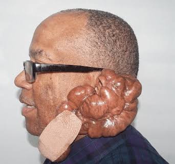
A keloid is a benign, firm, tumour-like fibrous swelling in the skin which arises in a scar. It is a form of excessive healing found only in humans.
Keloids are the result of an overgrowth of dense fibrous tissue that usually develops after healing of a skin injury. The tissue extends beyond the borders of the original wound, does not usually regress spontaneously and tends to recur after excision.
In contrast, hypertrophic scars are characterized by erythematous (difficult to see in the dark-skinned person),pruritic, raised fibrous lesions that typically do not expand beyond the boundaries of the initial injury and may undergo partial spontaneous resolution.
Hypertrophic scars are common after thermal injuries and other injuries that involve the deep dermis.
In local parlance, keloids are called bumps.
CAUSES OF KELOIDS
The aetiology is not understood; no specific gene or set of genes has been identified. However, the increased prevalence of keloids paralleling increased cutaneous pigmentation suggests a genetic basis, at least, a genetic linkage. Genetically,keloids have been linked with HLA-BI4, HLA-B2l , HLLBw16, HLA-Bw35 , HLA-DR5, HLA-DQW3 and blood group A.
Trauma to the skin, both physical(e.g.earlobe piercing,surgery) and pathological (e.g. acne, chickenpox),is the primary cause identified for the development of keloids.
The presence of foreign material, infection, haematoma or increased skin tension can also lead to keloid or hypertrophic scar formation in susceptic individuals.
Both dominant and recessive modes of inheritance have been described .
CLINICAL FEATURES
Keloids are found all over the world with a greater prevalence in women probably because of piercing of ears.
There is a preponderance in Africans(blacks) and this liability to keloid formation is higher in the darkly-pigmented racial groups.
They are commoner in dark-skinned individuals followed by Asians, then Caucasians, but rare in albinos of all races.
Although they occur in all age groups, they are hardy found in the newborn or elderly and the highest incidence is in those aged 10-30years. They occur in 5-l5% of wounds.
The areas most frequently affected are in the descending order the ear lobes, face, neck, lower extremities, breast,chest, back and abdomen. Keloid of the sole has been reported but it is rare.
It is raised above the skin level and extends beyond the margin of the wound. It continues to grow beyond a year and may then shrink a little.
The hypertrophic scar during the period of growth, which is less than a year, is raised above the skin, very vascular, painful and itchy; this growth period is then replaced by maturation and resolution when it becomes flat but broad.
A quiescent keloid may start growing again during pregnancy.
COMPLICATIONS
I. Ulceration from trauma.
2. Infection is common.
3. Malignant change. A fibrosarcoma may occasionally develop in a keloid.
4. Itching and tenderness.
TREATMENT
1. The treatment of keloid is unsatisfactory. No single modality is best for all keloids. Therapeutic measures include occlusive dressings, compression therapy,intralesional corticosteroid injections,excision, cryosurgery,radiotherapy and laser therapy. Excision alone has a recurrence rate of 50- 100%. Surgery alone should, therefore, be avoided.
2. Excision and radiotherapy: Surgical excision, followed by superficial irradiation (12Gy) started within 24hours after surgery, is not always successful. There is also the risk of late tumour induction.
3. Excision and triamcinolone (steroid) injection : Excision through the cdge of the keloid is followed immediately by injection of triamcinolone acetonide 20-80mg. The injection is repeated monthly for six months. This method is usually successful. Triamcinolone (kanalog) may be injected into small keloids without excision. Steroids impair the inflammmatory reaction responsible for the excessive production of collagen.
4. Triple therapy: This involves surgical excision of the keloid, followed by radiotherapy within 24hours and then triamcinolone injection. Triple therapy appears to give a better result than surgical excision and radiotherapy. There is risk of late tumour induction.
5. Compression therapy: Application of consistent controlled pressure to the wound decreases blood flow through the dermal vasculature and thereby prevents haphazard proliferation of fibroblasts and excessive collagen deposition.
The mechanical force applied to the wound alters the healing process resulting in a flat scar.
i) Button therapy on ear lobes: After excision of the keloid, two ordinary shirt buttons firmly sandwiching the lobule are threaded together and left in place for 6 months.
ii) Zinc oxide adhesive plaster is used for facial keloids.
iii) Ear clips can also be used.
6.i) Silicone cream/occlusive dressing: A firm occlusive dressing with a silicone cream containing 20% of silicone oil applied to the keloid has been found beneficial in some patients. It probably works by occlusion and hydration.
ii) Silicone gel: This can be applied to the keloid and held in place continuously using cellotape. The silicone is removed twice a day (morning and evening) and washed to get rid of sweat and re-applied . The mode of action is similar to that of silicone cream.
7. Recent innovation: New treatments for keloids and hypertrophic scars include intralesional interferon, 5FU,doxorubicin, bleomycin, verapamil, retinoic acid,imiquimod 5% cream, tacrolimus, tamoxifen, botulinum toxin and TGF-beta 3. More work needs to be done on these recent innovations before they can be used on all patients. Local creams for pruritus(itching) include lanoline and synalar, a triamcinolone cream. They are massaged after bath when the area is quite free of grease.
8. Hypertrophic scars are left alone as they show Spontaneous regression.
This article was adapted from Principles and Practice of Surgery by Badoe.
Read more on Wikipedia


